

Day 2 :
Keynote Forum
Francisco Capani
University of Buenos Aires, Argentina
Keynote: Degenerative modifications in synapses and related structures induced by experimental perinatal asphyxia in rat: Correlative light and electron microscopy studies.
Time : 10:15 -10:55

Biography:
I completed my PhD at the De Robertis Institute, School of Medicine-University of Buenos (UBA). I performed my postdoctoral studies at University of California San Diego, School of Medicine (UCSD-NCMIR) and Karolinska Institute, Department of Neuroscience. for 8 years focusing my research on cell biology of dendritic spines combining electron tomography and 3-D reconstruction techniques. When I returned to Argentina in 2007 I applied my broad experience in electron microscopy to study the mechanisms involved in pathophysiology of the perinatal asphyxia. Since 2018 I am director of Institute of Research in Cardiology, and Professor of Histology and Cell Biology, School of Medicine (UBA). I published 85 papers in reputed journals and has been invited in more than 14 international symposiums. I have been serving in the editorial in board of several scientific journal. I addition I was president of Interamerican Committe of Societies for Microscopy (CIASEM) for the period 2015-2017 .
Abstract:
Statement of the Problem: Diminish in the oxygen levels prompted short and long-term alterations in synapses and related structures that are related to neuronal dysfunction and death. Perinatal asphyxia (PA) is an obstetric complication produced by an impaired gas exchange that lead to neonatal mortality and is a determinant factor for neurodevelopmental disorders. Since pathophysiological mechanisms triggered by PA are not still totally unveiled, we investigated the changes in the cytoskeleton organization in the nervous tissue.
Methodology & Theoretical Orientation: For this study, we used a well-established murine model of PA. After one, 2, 4 and 6 months of severe PA (20 min) rats were sacrificed and their brains were analyzed by combining photooxidation, conventional electron microscopy, and 3-D reconstruction techniques.
Findings: After one month of PA, we found an increase in the F-actin staining in neostriatal and hippocampal dendritic spines together with some filopodia-likes structures, a typical embryonic type of spines in photooxidated tissue. In contrast, after second month of PA, spines were less consistent stained. In addition, we observed an increment of marker for neuronal and glial dysfunction such as GFAP, neurofilament and MAP-2. These modifications were more striking defined after 4 months of PA. After 6 months of PA post-synaptic densities (PSDs) in neostriatum were highly modified. Using three-D reconstructions and electron tomography we were able to find clear signs of degeneration in the asphyctic PSDs
Conclusion & Significance: Therefore, we hypothesize that the cytoskeletal changes induced by PA in the rat CNS could lead to the dramatic modifications in synapse and related structures that trigger neuronal damage. In addition, electron tomography, 3-D reconstruction and photooxidation contributed to dissect critical alterations generated by PA that are not easily displayed using conventional microscopic techniques. development.
Keynote Forum
Alice Zemljic-Harpf
University of California, USA
Keynote: Statin Toxicity: First report of atorvastatin, but not pravastatin, induced ultrastructural and functional changes in cardiac mitochondria
Time : 11:10 -11:50

Biography:
Dr. Alice Zemljic-Harpf has obtained her Medical Degree from the Medical University Graz, Austria in 1997. She conducted her postdoctoral studies in molecular cardiology at the Cedars-Sinai Medical Center Los Angeles (1999-2000), UCLA (2000-2003) and UCSD, School of Medicine. She is the director of The Cardiovascular Physiology and Imaging Unit at the Cardiac/Neuro Protection Laboratories at UCSD, Department of Anesthesiology. She has published 24 papers in high-impact pier reviewed journals (Circulation, Circulation Research, Glia, Cerbarl Cortex, The FASEB Journa, Heart Rhythm, The American Journal of Pathology, etc.). Her most recent work on atoravstatin induced adverse events gains international attention.
Abstract:
Background: Statins are amongst the most widely prescribed drugs to reduce LDL-cholesterol for the treatment of cardiovascular disease. Approximately one in five people in the United States between the ages of 45 and 75 take a statin. Like all drugs, statins can cause harmful side effects, such as muscle pain/weakness (statin myopathy), fatigue, nerve pain, and cognitive impairment. Because statin-induced myopathy is known to be associated with reduced oxidative phosphorylation in mitochondria of skeletal muscle we hypothesized that similar effects would occur in cardiac muscle.
Methods and Results: When male mice underwent atorvastatin and pravastatin administration per os for up to 7 months, only long-term atorvastatin, but not pravastatin administration induced: 1) elevated serum creatine kinase, 2) swollen, misaligned, size variable, and disconnected cardiac mitochondria, 3) altered ER-structure, 4) repression of mitochondrial and endoplasmatic reticulum related genes, and 5) 21% increased mortality in cardiac-specific vinculin knockout- mice.
Neonatal cardiac ventricular myocytes were treated with atorvastatin and pravastatin for 48hours. Both statins induced ER-stress, but only atorvastatin: 1) inhibited of ERK1/2T202/Y204, AktSer473 and mTOR signaling, 2) reduced protein abundance of caveolin-1, dystrophin, epidermal growth factor receptor and insulin receptor-β, 3) decreased RhoA activation, and 4) induced apoptosis. In cardiomyocyte-equivalent HL-1 cells atorvastatin, but not pravastatin, reduced mitochondrial oxygen consumption.
Conclusion and Clinical Implication: Skeletal muscle biopsies from patients with statin myopathy show increased lipid storage and alters mitochondrial structure. We are the first to demonstrate in vivo that long-term atorvastatin administration altered cardiac ultrastructure, a finding with important clinical implications.
- Molecular Cytopathology | Urinary Cytology | Digital Cytopathology
Session Introduction
Aleksandra Zuraw
Charles River Laboratories Montreal, Canada
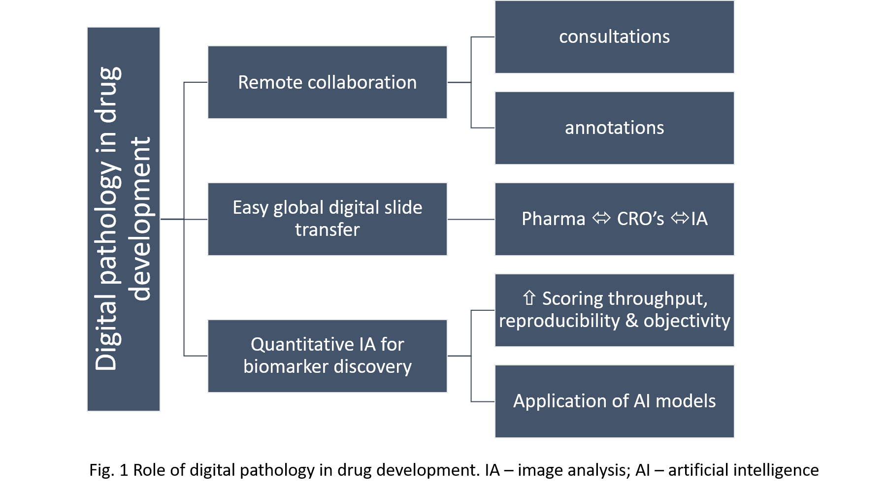
Title: Role of digital pathology in drug development process

Biography:
Aleksandra Zuraw is an ACVP board-certified veterinary pathologist with extensive experience in applying digital pathology to drug development. She is currently a Veterinary Pathologist at Charles River Laboratories Montreal, Canada. In addition to histopathological evaluation of pre-clinical animal studies, she has extensive expertise in digital image analysis powered quantification of immune-oncology tissue biomarkers and has worked closely with computer scientists and translational scientists to provide support for the drug development process. She obtained her DVM from the Wroclaw University of Environmental and Life Sciences, Poland and her PhD at the Freie Universität in Berlin, Germany.
Abstract:
Digital pathology is the process of performing pathology services using computers and digital images instead of microscopes and glass slides. With the increasing capabilities of pharmaceutical companies and contract research organizations to rapidly perform whole slide imaging and convert glass slides into digital images, there is a great potential to unlock the benefits of this technology for the drug development process. Pathologists are crucial members of drug development teams and are engaged in every step of the process, contributing significantly to the discovery, preclinical and clinical phases. Implementation of digital pathology workflows within and across organizations empowers them and benefits the drug development process in multiple ways. Often pathologists involved in pharmacological studies are geographically dispersed. The use of digital pathology allows them to communicate fast and perform slide consultation in real time regardless of location, which increases the efficiency of their work. Multiple pathologists can view, annotate and comment on the same slide simultaneously. The digitization of slides enables image analysis-powered quantitative measurements of biomarkers, lesions and abnormalities, which increases the throughput, reproducibility and objectivity of pathologists’ scoring. As tissue research is cross-disciplinary, access to digital pathology for different groups involved in drug development, especially the DVM and MD pathologists, helps them work more collaboratively and better understand their contributions. This technology is still considered novel, and in many institutions, its implementation is met with skepticism. Nevertheless, making it accessible and user-friendly for pathologists, standardizing it and applying it broadly across organizations, will undoubtedly accelerate and advance drug development.
Jamshid Abdul-Ghafar
French Medical Institute for Mothers and Children (FMIC), Afghanistan
Title: Compression of epidermal growth factor receptor (EGFR) gene expression in the malignant mesothelioma of Korean patients using fluorescence in situ hybridization (FISH), immunohistochemistry and gene mutation

Biography:
Dr. Jamshid Abdul-Ghafar has his expertise in Histopathology and Cancer research. He has done his M.D. degree in Afghanistan. He has joined Yonsei University, Wonju College of Medicine in South Korea for his Ph.D. in the Department of Pathology. He is currently working at French Medical Institute for Mothers and Children (FMIC) in Kabul, Afghanistan. Besides being a Pathologist, he has involved in Research and Academic works as well. He is working as Administrator of Postgraduate Medical Education and Program Director for Pathology residents. He has published more than 15 articles in different international science-indexed journals.
Abstract:
Introduction: Malignant mesothelioma (MM) is well known as aggressive cancer which has the resistance to usual treatments and show poor prognosis. Little has been reported regarding the effective therapeutic methods of MM. Recently, epidermal growth factor receptor (EGFR) gene has been introduced that it has important role in the biologic features and tumor growth of the pleural MM, and the novel therapeutic agent such as EGFR tyrosine kinase inhibitor attempt to treatment of MM. We evaluated the expression of EGFR gene using Immunohistochemical stains (IHC), fluorescence in situ hybridization (FISH), and EGFR gene mutation in cases of MM of Korean patients. Methods: We reviewed clinicopathologic findings for 44 cases. The mean age of cases (male 32, female 12) was 59 years old (19-79). Most cases were occurred in the pleura (38 cases, 83.4%) and 6 cases (13.6%) from the peritoneum. The IHC for EGFR antibody for 29 cases, FISH analyses for 17 cases and detection of EGFR gene mutation for 36 cases were performed. The histologic subtypes were epithelioid in 31 (70.5%), biphasic in 5 (11.4%), desmoplastic in 3 (6.8%), and other variants in 5 (11.4 %). Results: On the result of IHC stains for EGFR, 7 cases (24.1%) showed positive expression. According to the EGFR FISH analysis only 2 cases (11.8%) revealed a positive gene copy number gain of EGFR (high polysomy), both cases were in IHC-positive group. Thirty-five cases of MM did not show any EGFR mutation. EGFR mutation was found in only one case of MM on exon 21, L861. Conclusion: EGFR overexpression is relatively common in epithelioid type of MM. The protein expression of EGFR is not significantly related to a gene copy number gain and nor related to EGFR mutation. However, the further studies to the larger group in future are expected.
Akinwale Damilola Ayodeji
Ladoke Akintola University of technology, Nigeria
Title: Association between secretor status and hepatitis B infection

Biography:
Akinwale Damilola has completed his BSc. in medical laboratory sciences at the age of 27 years from Ladoke Akintola University. He is an intern medical laboratory scientist at Adeoyo maternity teaching hospital, yemetu, Ibadan, Oyo State, Nigeria.
Abstract:
Hepatitis B is an infectious disease caused by the hepatitis B virus (HBV) which affects the liver. It can cause both acute and chronic infections. The aim of this study is to investigate the association between secretor status and Hepatitis B infection. Secretor status was determined by agglutination inhibition method. This study was carried out among 200 subjects, of whom 100 were hepatitis B patients and 100 apparently health subject that served as control. Of the 100 hepatitis B patients, 33(33.0%) were males and 67(67.0%) were females. Of the 33 males, 19 were secretors and 14 were non-secretors while 45 and 22 of the females were secretors and non-secretors respectively. There is no significant association between secretor status and sex (2=1.7, df =1, p= 0.192). This study shows that there is no significant association between secretor status and hepatitis B.
Hira Kareem
Services Institute of Medical Sciences, Pakistan
Title: Glomerular changes in kidney of albino rats after using copper diet: A biological study

Biography:
Hira Kareem has completed her post-graduation from the University of Health Sciences Lahore at the age of 28 years. Currently, she is serving as a demonstrator in Services Institute of Medical Sciences, Lahore. She has published three medical papers in reputed journals.
Abstract:
Aim: An experimental study to evaluate the effects of copper metal on renal glomeruli. This study was conducted from May to November 2013 in the Pathology Department of the University of Health Sciences, Lahore.
Methods: 20 albino rats were selected and divided into two groups. The control group 1 was fed on standard pellet rodent diet and tap water. The experimental group 2 was fed 0.5mg/kg of body weight for 18 weeks on alternate days; the dose was calculated according to the body weight. Rats were sacrificed at the end of the experiment and the kidneys were removed for processing and H&E staining for routine histological study. Glomerular changes observed by microscope and statistically analyzed using the Chi-square test with p<0.05.
Results: Results showed that glomerular changes in terms of mesangial cell proliferation by using copper in the diet of rats. When the control group 1 on a normal diet was compared with the experimental group 2, a statistically significant difference (p<0.05) was noticed.
Conclusion: It is therefore concluded that copper in the diet affects the glomeruli in terms of mesangial cell proliferation, adhesion and crescent formation in kidneys of albino rats.
Sohair M Sokkar
Cairo University, Egypt
Title: Comparative pathological studies on Cryptococcus neoformans in experimental animals
Biography:
Abstract:
A comparative study was done to differentiate the pathogenicity and the histopathological lesions in guinea pigs and mice either male or female infected with either field isolate or standard strain of C. neoformans. Also, a comparison was done to differentiate the lesions by the different routes of infection. Seventy adult male and female guinea pigs and seventy adult male and female mice were used. The dose of injection was 2x10⁷CFU/ml. Four animals from each inoculated group and two untreated control animals were slaughtered every 10 days until the end of the experiment. Tissue specimen from brain, lung, liver, spleen, and kidney were collected for the histopathological examination, reisolation of C. neoformans and estimation of macrophage phagocytic activity (alveolar macrophage in guinea pigs, peritoneal macrophage in mice). The alveolar macrophages in guinea pigs and the peritoneal macrophages in mice were able to engulf the yeast. The most prominent postmortem lesions were congestion of the brain meninges and the lung showed diffuse pneumonia. In guinea pigs, there was severe granulomatous pneumonia with systemic dissemination of the infection. In mice inoculated intradermally, there were cutaneous lesions. The most prominent postmortem lesions were multiple granulomas in the lung with severe pneumonia, spleen and liver showed mucoid nodules. The cutaneous lesions appeared as elevated, roughened, hairless plaques. The lesions either after infection with the field isolate or the standard strain were nearly the same but those of the field isolate was more severe. The sex (either male or female) played no role.