Day 1 :
Keynote Forum
Vinod B. Shidham
Wayne State University School of Medicine, USA
Keynote: Cellblochistry: Chemistry & Art of Cell Block making
Time : 10:00-10:40

Biography:
Dr. Vinod B. Shidham is a certified Anatomic and Clinical Pathologist and Cytopathologist by American Board of Pathology. His spectacular career started in Nagpur (Maharashtra State). After serving in academia in India up to the level of Professor of Pathology at Grant Medical College and JJ Group of Hospitals in Mumbai till 1985, he left for middle east and then to USA. Currently, he is Professor of Pathology at Wayne State University School of Medicine, Karmanos Cancer Center, & Detroit Medical Center, Detroit, MI in USA. He is Vice-chair- Anatomic Pathology and Director of Cytopathology, Cytotechnology School, Cytopathology fellowship training program, GI pathology fellowship, and Pathology Residency Training Program.
Dr. Shidham is a strong proponent of ‘Open access’ philosophy in academic publications. He is editor-in-chief of ‘CytoJournal” which is a peer-reviewed, PubMed indexed open access journal publishing high-quality papers in cytopathology. He is editor of upcoming monographs on ‘EUS-FNA of pancreas’ and ‘FNAB procedure’ as CytoJournal Monograph & Atlas Series on timely topics.
Abstract:
CellBlochistry is science to study the art and chemistry of cell block making. It involves conglomeration of minute tissue fragments and isolated cells / small cell groups in specimens collected or submitted as suspensions. This conglomeration is processed as paraffin embedded tissue sections with benefits of long term archival and other benefits comparable to surgical pathology biopsy specimens for various elective ancillary tests.
Variety of samples that may be ‘Cell Blocked’ include FNA needle rinses, body fluids, exfoliated cells similar to the cervical cytology specimens, different brushings such as bronchial, bile duct etc, various curettages including endocervical curettings, scrapings of cytology smears (stained or unstained), or any other cytology specimen with micro-fragments. In addition, research samples including scrapings of surface grown cell cultures and specimens from animal experiments (comparable to clinical samples) or comparable veterinary specimens.
The role of cell blocks is already established but it’s significance is increasing further due to their enhancing role with recent advances in molecular pathology in modern medicine. However, variable complexity on both, scientific and skill fronts, introduces many challenges with suboptimal quantitative and qualitative outcomes.
The current keynote presentation will cover the limitations related to various alternatives available for cell block preparation with solutions to overcome reasons for suboptimal cell blocks and cell block sections. Leading factors responsible for such interferences include fixation and processing. Immunocytochemical evaluation as most frequent indication for cell blocks is facilitated by applying Subtractive Coordinate Immunoreactivity Pattern( SCIP) approach.
Additionally, other ancillary testing which can be performed on blocks include molecular pathology studies, In situ hybridization studies (FISH, CISH), Histochemistry (stains for organisms, various tissue structures/components), and other evolving technologies such as mass spectrophotometry, proteomics, etc.
Keynote Forum
Hamed A. Benghuzzi
Department of Diagnostic and Clinical Health Sciences, SHRP, UMC
Keynote: Histopathological evaluation of fibrous tissue surrounding bioceramic delivery systems in sheep and rodent models
Time : 10:55-11:35

Biography:
Dr. Benghuzzi is a Professor at the University of MS Medical Center. He is known nationally and internationally as a pioneer in Ceramic Drug Delivery Systems. He has over 250 PubMed indexed articles and over 700 abstracts detailing the release characteristics of various biologicals from the bioceramic carriers. He has trained more than 40 PhD students who are actively involved in academic careers. He has mentored students at all levels (from high school, undergrad, grad, post doc and faculty). He has served as a mentor for residents and faculty on more than 10 funded grants. He has been in research leadership roles in many organizations such President of the Academy of Surgical Research, Vice President of the Rocky Mountain Bioengineering Society, President of MAS, Academy’s Executive Director, and also organized and chaired several regional, national and international society programs. He has also served on numerous NIH special emphasis panels including R-25, K01, KO8, T-35, and the P-60 center grants. In addition, he has received numerous awards from various organizations during his career. A few of his awards included: (1) The Presidential Award from the RMBS, (2) Presidential Award from SEM International, (3) the Endocrine’s Society Outstanding Investigator Award, (4) MAS Contribution to Science Award, (5) The MAS Dudley Peeler Award, and (6) HEADWAE Award, (7) C. Hall Award, Outstanding Contribution to Biomedical Engineering (32nd SBEC), and (8) ISCM Excellence Award from the International Society for Ceramics in Medicine.
He was invited as a keynote/plenary to speak at state, national and international levels including recent invitations in France, Italy, Spain, Greece, China, Poland, Dubai and Canada. He is a fellow of the American Institute for Medical and Biological Engineering (AIMBE) as well as an International Fellow of Biomaterials Science and Engineering (FBSE).
Abstract:
The major challenges faces vast majority of orthopedic and dental implants are: (i) biocompatibility, (ii) resorbability and (iii) maintenance of mechanical strength. Several studies conducted in our laboratories have documented the effectiveness of various ceramic drug delivery systems (CDDS) in regulating fertility in females as well as males. It was observed that the mode of sustained delivery of reproductive hormones from CDDS was governed and regulated by the development of capsular tissue surrounding the implantable CDDS. This investigation was specifically designed to correlate the thickness of fibrous capsule and the various histopathological components that are seen in the fibrous capsule surrounding ALCAP, HA, and TCP ceramics at the S/C and I/P implantation sites in small (rats) and large (sheep) animals. Male albino rats and castrated adult rams were utilized for these studies. All experimental animals were implanted either S/C or I/P with ALCAP, HA, and TCP ceramics delivery devises loaded with 40 mg testosterone. At 90 days post- implantation, the animals in all groups were euthanized and the fibrous tissue surrounding the ceramic devices and internal organs were harvested. After routine histological processing, sections of tissue were stained with hematoxylin and eosin as well as special stains and evaluated using light microscopy. Analysis of the data revealed the followings: 1) development of fibrous tissue around the implants did not show any significant difference in small or large animals, 2) capsular tissues retrieved from S/C site induced less fibrous tissue formation compared to I/P site in both models, 3) the presence of proinflammatory cells such as macrophages, neutrophils, fibroblasts, and vascularity, was found to be statistically different among the S/C implanted CDDS groups (p<0.01) compared to I/P implantation site, and 4) regardless of animal model, the ease of fibrous capsule thickness were ALCAP> HA>TCP, respectively. Results from these studies provided very important outcomes in which can be utilized to predict the longevity of the physical strength of CDDS and the duration of delivery profiles. The clinical impact of this investigation stems in the ability to develop biocompatible and effective delivery systems that ultimately will lead to fertility regulation.
Keynote Forum
Noeme Sousa Rocha
UNESP: - Portal of the State University of São Paulo, Brazil
Keynote: Equine melanoma- Anatomoclinical expression and biological behavior
Time : 11:35-12:15

Biography:
Noeme Sousa Rocha was graduated in Veterinary Medicine from State University of Maranhão (1989), completed Masters in Pathology from São Paulo State University (1994) and Ph.D. in Pathology from São Paulo State University (1998). She is currently an Associate Teacher of São Paulo State University, Brazil and she has experience in the area of veterinary medicine, with emphasis on animal pathology anatomy, acting on the following topics: veterinary, cytopathology, pathology, cancer and histopathology. She is also an Associate Member of the International Academy of Pathology.
Abstract:
Equine Melanoma – Anatomoclinical Expression and Biological Behavior: Immunohistochemistry techniques allow visualise for the visualization of specific markers relevant to the diagnosis and prognosis of neoplasms, since them they are characteristic of particular cellular events. In horses, variations in proliferation and adhesion markers expression were associated with the degree of melanoma malignancy. Although this tumor is not a recent entity, researches research has not accompanied the evolution of anatomoclinical diagnostic techniques. Due to it, we identified possible relationships between anatomoclinical expression and biological behavior, correlating them with the degree of tumor malignancy. For this, horses with the diagnosis of melanoma confirmed by cytopathological and histopathological exams were selected from the Archive of the Veterinary Pathology Department, without predilection for race, gender or age. Cytopathological slides were analyzed for the presence of atypical melanocytes, while those stained with HE were evaluated according to micrometric criteria of "Breslow". Furthermore, we analyzed the presence of lymphocytic infiltration, angiolymphatic invasion, necrosis and mitosis. Avidin-Biotin-Peroxidase technique was used for immunolabeling reactions that identify S-100, Ki-67, E-cadherin and CD44. Thus, cell receptors of the proliferative phase, loss of adhesion, migration and evasion were observed. Density of the immunolabellated immunolabeled cells of each antibody were determined by five fields of greatest magnification under optical microscope. The results were submitted to statistical analyzes analysis. We intend contribute to a better understanding of the biological behavior of this tumor, in order to demonstrate that a quickly carried out diagnosis containing several criteria of malignancy has significant importance in the patient’s prognosis.
- Diagnostic Cytopathology | Cancer Cytopathology | Histopathology | Exfoliative Cytopathology | Surgical Pathology | Fine Needle Aspiration Cytology
Session Introduction
Michelle Tucci
University of Mississippi Medical Center, USA
Title: Unraveling the mechanism of actions for thymoquinone and EGCG on cancer cells
Time : 12:15-12:45

Biography:
Michelle Tucci, Professor of Anesthesiology at the University of Mississippi Medical Center, has been heavily involved with teaching, research and services at the institutional, state, national and international levels. After completing undergraduate training at Seton Hill University, she completed a master’s degree in Biology at the University of Dayton. Following her move to Mississippi, she completed her PhD in pharmacology in 2000. Aside from her work supervising and overseeing resident research in biomedical sciences, she has also mentored and supervised a number of undergraduate and graduate students from diverse disciplines. She has served on 38 doctoral dissertation committees (chair and member/see CV), has published over 209 full journal publications, and presented 327 abstracts over her career at state, regional, national and international meetings (Italy, France, Spain, Greece, Canada, and China). She served in leadership role at various societies such as Director and program chair at the Rocky Mountain Biomedical Engineering Society; Chair of Pathology Implant SIG at the Society for Biomaterials, Program Chair at Southern Bioengineering Conference to name a few. She served/serving in editorial boards in several journals as well as member of various NIH special review panels. She secured intramural research funding from American Cancer Society, Orthopedic Trauma Association, IsoTish Inc, Inplex Inc, Enzymatics and mentored several orthopedic residents training grants. She has tirelessly served the several biomedical engineering organizations with distinction in a number of capacities including division chair and vice-chair, poster session chair, the board of directors, and most recently as editor of the JMAS. Previously, she has been recognized for her work and services by many societies such Teacher of the year, Peeler Dudley service award, Walker outstanding research awards.
Abstract:
Natural products like epigallocatechin gallate (EGCG) and Thymoquinone (TQ). EGCG is considered to be responsible for most of the healthy benefits associated with green tea. In particular, EGCG is a polyphenol, which helps with the production of certain proteins and alkaloids. TQ is a compound derived from the black seeds called Nigella sativa. TQ has been used for more years as a medicinal herb to fight disease and boost immunity. Our recent findings have shown that EGCG and TQ to be effective in interrupting the growth of many different types of cancer cells. The exact mechanism of action are currently unknown. We have investigated both compounds for their effectiveness in reducing cancer cell loads in numerous cell lines and each compound appears to an IC50 dose that is cell line specific as well as targets different signaling pathways. Overall, functional and histopathological evaluation of both compounds may be more effective in combination with other chemotherapeutic agents to ultimately arrest the invasiveness of neoplastic cells.
Punam Prasad Bhadani
All India Institute of Medical Sciences, India
Title: Diagnostic accuracy of fine needle aspiration cytology of thyroid lesions and its histopathological correlation

Biography:
Punam Prasad Bhadani has done her medical graduation (M.B.B.S) in 1991 from Darbhanga Medical College, Bihar, India, and post-graduation (MD) in Pathology from Patna University, India in the year 1999. She worked as Assistant & Associate Professor at B.P. Koirala Institute of Health Sciences, Dharan, Nepal, Associate Professor at Government Medical College Haldwani, Uttarakhand, India, and Professor at School of Medical Sciences, Sharda University, Greater Noida, India.
Presently she is Addl. Professor & Head, Department of Pathology, All India Institute of Medical Sciences, Bihar, India. She is the Editorial Board Members in two journals and published 60 paper and author of two books.
Abstract:
Fine needle aspiration cytology (FNAC) is a simple, cost-effective and widely used procedure for thyroid lesions. A study is undertaken to correlate the FNAC findings with histopathology in a spectrum of thyroid lesions. To determine the diagnostic accuracy of FNAC, it was compared with the histopathology to highlight its usefulness. This retrospective study was done in an upcoming tertiary care hospital of the eastern state, Bihar of India. This study was done for evaluation of FNAC findings for Thyroid lesions in a span of three years from 2014 to 2017. Records of 356 cases of thyroid lesions were studied, who underwent FNAC for thyroid lesions. Mean age of the patient was 42 years with a female to male ratio of 4.1:1. The cytological results were classified as inadequate, benign, suspicious, and malignant. According to FNAC diagnostic criteria, 22 cases (6.2%) were diagnosed as inadequate, 309 cases (86.8%) benign, 10 (2.8%) suspicious and 15 (4.2%) malignant for diagnosis. In the study, a cytohistological correlation was done in 42 cases. In which they were classified as non-neoplastic (benign) and neoplastic (malignant) lesions. Out of 42, 33 were classified as benign and 09 as malignant lesions. The sensitivity, specificity, and diagnostic accuracy were 71.4%, 100%, and 94.8%, respectively.
CONCLUSION:
FNAC is a safe, simple, highly accurate, economical and universally accepted tool for the evaluation and management of thyroid lesions. FNAC also helps to avoid unnecessary surgical intervention in patients of a benign pathology of the thyroid gland. This study has shown that it has high specificity and accuracy and reinforces the need for its continuous practice in the management of thyroid lesions.
Distribution of benign (a), suspicious (b) and malignant(c) cases on cytology
|
Different thyroid lesions |
Frequency |
Percentage (%) |
Lesions (N = 309)
|
|
|
|
Colloid goiter |
223 |
72.2 |
|
Hyperplastic/toxic nodule |
15 |
4.9 |
|
Hashimoto thyroiditis |
67 |
21.7 |
|
Subacute thyroiditis |
02 |
0.6 |
|
Suppurative thyroiditis |
02 |
0.6 |
|
b) Suspicious cases Lesions (n = 10)
|
|
|
|
Follicular neoplasm
|
06 |
60 |
|
Hurthle cell neoplasm |
04 |
40 |
|
c) Malignant cases Lesions (n = 15)
|
|
|
|
Papillary carcinoma |
09 |
60 |
|
Medullary carcinoma |
01 |
6.7 |
|
Anaplastic carcinoma |
04 |
26.6 |
|
Lymphoma |
01 |
6.7 |
Daniela Cristina dos Santos
São Paulo State University, Brazil
Title: Peritoneal malignant mesothelioma in young male patient: Ten years of evolution

Biography:
Daniela Cristina dos Santos was graduated in Medicine from São Paulo State University (2005), completed Medical Residency in Pathology (2009) from São Paulo State University, Masters in Pathology from São Paulo State University (2011) and completed Ph.D. at the age of 38 years from São Paulo University in Pathology. She is currently Medical Assistant of the Department of Pathology of São Paulo State University, Brazil. She has experience in the area of Cytopathology, Histopathology, Fine Needle Aspiration Cytology (FNAC) in cancer diagnosis, with emphasis on Pathology Transplantation and Nephropathology. She is also supervisor of the Division of Postmortem Inspection.
Abstract:
Peritoneal Malignant Mesothelioma in Young Male Patient: Ten Years of Evolution: Epithelioid peritoneal malignant mesothelioma in patient male, young, with absence of history of asbestos exposure and with supraclavicular lymph node metastasis. Abdominal ultrasonography and computed tomography showed large amount of ascites into peritoneal cavity. On gross examination, at laparoscopy abdominal, gray nodules with variable size (1 to 6 milimeters) and morphology with nodules coalescent and plaques involving peritoneal and liver surface are observed. Fine Needle Aspiration (FNA) in lymph node with Giemsa and Papanicolaou (PAP) staining show, on microscopic examination, large cell clumps with cuboidal mesothelial cells with increased nuclear to cytoplasm ratio and unclear cell borders, concentric round nuclei, conspicuous nucleoli and sparse mitotic figures. The stroma is fibrotic and myxoid. Tumor cells were positive for calretinin (strong nuclear and cytoplasmic expression), D2-40, EGFR, cytokeratin 5/6 and WT1, but were negative for MOC31, CEA, TTF-1, and Ber-EP4. We expected to contribute in the diagnosis identification with an unusual peritoneal mesothelioma in young male patient with clinic evolution more than 10 years.

Biography:
Abstract:
The occurrence of primary neoplasia of the central nervous system is associated with broad impairment of the host immunocompetence. In the present studies an ultrastructure analysis of peripheral blood lymphocytes of gliomas patients were carried using Transmission Electron Microscopy and Scanning Electron Microscopy. Under SEM lymphocytes presented smooth surface with loss of microvilli.These cells were referred as Bald Cells.Similar loss of microvilli and altered cell membrane was observed under.TEM. Generally, a low level of immunostaining (CD4+) was predominant in patient group and the Bald Cell failed to bind any of the monoclonal antibodies.It is hypothesised that activated T cells get activated by the tumor associated antigen releasing in peripheral blood via break down of blood brain barrier.An elevated level of soluble IL-2 receptors observed in serum confirm the shedding phenomenon of surface receptors by activated T Cells.
The loss or shedding of phenotypic determinants from the surface of lymphocytes leads to either a low level of immunostaining or complete failure of staining by the monoclonal antibodies.The apoptosis phenomenon, subsequently leads to death of cells. This may be one of the factors responsible for the significant CD3/CD4 lymphopenia observed in gliomas patients.
However, revival of Bald Cells is crucial to restore the immunity.
Present study carried out using immuno histopathological technique (PAP &ABC methods with commercially procured monoclonal antibodies).The blood lymphocytes were also viewed under TEM before and after immunostaining.
Sandhya Sundaram
Sri Ramachandra Medical College & Research Institute, India
Title: Over expression of C326 (EpCAM) in carcinoma breast and its clinical correlation

Biography:
Sandhya Sundaram is a senior pathologist with 21 years of experience in practice of Pathology. She completed her MD &DNB in pathology and Postdoctoral certificate course in Laboratory Medicine. She has more than 50 publications in reputed journals and several national research grants. Her basic area of interest is oncopathology with a keen interest in Breast pathology and Genitourinary and gastric Pathology. She is keenly interested in Morphological, Molecular Pathology and Molecular genetics of solid tumours and infections associated with malignancies and their molecular characterization. She has also worked in fungal infection in collaboration with Microbiology Department
Abstract:
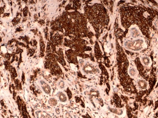
Statement of the Problem: The epithelial cell adhesion molecule (EpCAM) is a type I transmembrane protein of 314 amino acids that is localized to the basolateral membrane in the majority of normal epithelial tissues.EpCAM has been demonstrated to be a calcium-independent homophilic cell adhesion molecule. Recent studies have also demonstrated a role for EpCAM in cell signaling and carcinogenesis. EpCAM is overexpressed in a variety of epithelial tumours such as gastric cancer, oesophageal cancer and prostate cancer The main purpose of the study was to determine the expression pattern of EpCAM across molecular subtypes of carcinoma breast in the South Indian population and to correlate its expression with other clinical parameters. immunohistochemical studies were performed on 50 cases of breast carcinoma of different molecular subtypes. This included luminal A, Luminal B, Her 2 Neu & triple-negative subtypes. The expression was scored using the standard scoring system A correlation was drawn with detailed clinicopathological annotation and available outcomes data. We observed strong EpCAM expression in 42 out of 50 cases. EpCAM expression varied significantly in the different intrinsic subtypes. Expression pattern was membranous.EpCAM expression is higher in TNBC & Her 2 Neu type of Breast Carcinoma. Higher expression was seen in node-positive tumours. EpCAM expression has critical and important implications in subsets who would benefit from targeted therapies.
Images: Breast carcinoma cells showing strong EpCAM IHC
Positivity compared to normal glands which show very faint staining
Biography:
Enas Abdel-Hafez from Assiut University, Egypt
Abstract:
The aim of the current study was to estimate the metacercarial infection in Ruby-Red-Fin Shark, (Rainbow Shark), Epalzeorhynchos frenatum (Teleostei: Cyprinidae). Samples of fish were commercially purchased from an ornamental shop in Asyut city, Egypt and brought to the laboratory. Fish samples were used for light histological analysis. All fish measured and deeply anesthetized with benzocaine (4 mg/L). Fish were ranging from 10 to 12 cm in standard body length. Using histological examination, we identified a heterophyid parasite metacercaria; Ascocotyle (Ascocotyle) tenuicollis Price, 1935. The metacercarial cyst was located inside the cartilaginous support of the gill arches which were surrounded by thick fibrous perichondrium. Cartilage enlargement occurs as a result of invaginations of the perichondrium into the cartilage mass and proliferation of the chondrocytes progeny.
Ahmed El-Habashi
Tulane University Hospital, New Orleans, USA
Title: Thyroid lesions fine needle aspiration cytology: Comprehensive cytomorphology update reporting, diagnostic pitfalls and interactive case presentation

Biography:
Prof. DR. Ahmed El-Habashi has completed his MD, PhD at the age of 40 years from Cairo University and Tulane University in a channel system scholarship program. He got post-doctoral fellowship at Groningen University Hospital, The Netherlands at 1998. He is the past- director of pathology department and cytology division,, National Cancer Institute, Cairo University and now he is working in the capacity of Professor of Pathology. He got the International Board of Cytopathology from International Academy of Cytopathology at 2007. He is an international cytology speaker and he conducted many Cytopathology workshops in Egypt and Arab countries as well as in Canada. He has published more than 40 papers in reputed journals and has been serving as a reviewer for more than three reputed Journals.
Abstract:
FNAC is recommended as a routine prime investigation in the work-up of the solitary cold thyroid nodule. Requirements for accurate diagnosis are adequate material, optimal smearing, proper staining and experience. The accuracy of FNAC in thyroid lesions is over 90%, with sensitivity of malignancy diagnosis almost 100%, false negative is about 10% and false positive is 1-3%. The author in this workshop will demonstrate cytomorphologic features of majority of diagnostic entities in thyroid lesions FNA together with an emphasis on potential diagnostic pitfalls. The updated Bethesda System (TBS, 2017) for thyroid lesions FNA cytology reporting will be discussed with its clinical significance and management guidelines. The end of the talk will include interactive case presentations to ensure that the audience will achieve the basic ILOs objectives of the workshop.
Francisco Capani
University of Buenos Aires, Argentina
Title: Degenerative modifications in synapses and related structures induced by perinatal asphyxia: Correlative ligth and electron microscopy studies.

Biography:
I completed my PhD at the De Robertis Institute, School of Medicine-University of Buenos (UBA). I performed my postdoctoral studies at University of California San Diego, School of Medicine (UCSD-NCMIR) and Karolinska Institute, Department of Neuroscience. for 8 years focusing my research on cell biology of dendritic spines combining electron tomography and 3-D reconstruction techniques. When I returned to Argentina in 2007 I applied my broad experience in electron microscopy to study the mechanisms involved in pathophysiology of the perinatal asphyxia. Since 2015 I am vice director of Institute of Research in Cardiology, and Professor of Histology and Cell Biology, School of Medicine (UBA). I published 85 papers in reputed journals and has been invited in more than 14 international symposiums. I have been serving in the editorial in board of several scientific journal. I addition I am president of Interamerican Committe of Societies for Microscopy (CIASEM)
Abstract:
Statement of the Problem: Diminish in the oxygen levels prompted short and long-term alterations in synapses and related structures that are related to neuronal dysfunction and death. Perinatal asphyxia (PA) is an obstetric complication produced by an impaired gas exchange that lead to neonatal mortality and is a determinant factor for neurodevelopmental disorders. Since pathophysiological mechanisms triggered by PA are not still totally unveiled, we investigated the changes in the cytoskeleton organization in the nervous tissue.
Methodology & Theoretical Orientation: For this study, we used a well-established murine model of PA. After one, 2, 4 and 6 months of severe PA (20 min) rats were sacrificed and their brains were analyzed by combining photooxidation, conventional electron microscopy, and 3-D reconstruction techniques.
Findings: After one month of PA, we found an increase in the F-actin staining in neostriatal and hippocampal dendritic spines together with some filopodia-likes structures, a typical embryonic type of spines in photooxidated tissue [Fig 1 A). In contrast, after second month of PA, spines were less consistent stained. In addition, we observed an increment of marker for neuronal and glial dysfunction such as GFAP, neurofilament and MAP-2. These modifications were more striking defined after 4 months of PA. After 6 months of PA post-synaptic densities (PSDs) in neostriatum were highly modified. Using three-D reconstructions and electron tomography we were able to find clear signs of degeneration in the asphyctic PSDs (Fig 1 B and C).
Conclusion & Significance: Therefore, we hypothesize that the cytoskeletal changes induced by PA in the rat CNS could lead to the dramatic modifications in synapse and related structures that trigger neuronal damage. In addition, electron tomography, 3-D reconstruction and photooxidation contributed to dissect critical alterations generated by PA that are not easily displayed using conventional microscopic techniques.
Noeme Sousa Rocha
UNESP: - Portal of the State University of São Paulo, Brazil
Title: Equine melanoma- Anatomoclinical expression and biological behavior

Biography:
Noeme Sousa Rocha was graduated in Veterinary Medicine from State University of Maranhão (1989), completed Masters in Pathology from São Paulo State University (1994) and Ph.D. in Pathology from São Paulo State University (1998). She is currently an Associate Teacher of São Paulo State University, Brazil and she has experience in the area of veterinary medicine, with emphasis on animal pathology anatomy, acting on the following topics: veterinary, cytopathology, pathology, cancer and histopathology. She is also an Associate Member of the International Academy of Pathology.
Abstract:
Equine Melanoma – Anatomoclinical Expression and Biological Behavior: Immunohistochemistry techniques allow visualise for the visualization of specific markers relevant to the diagnosis and prognosis of neoplasms, since them they are characteristic of particular cellular events. In horses, variations in proliferation and adhesion markers expression were associated with the degree of melanoma malignancy. Although this tumor is not a recent entity, researches research has not accompanied the evolution of anatomoclinical diagnostic techniques. Due to it, we identified possible relationships between anatomoclinical expression and biological behavior, correlating them with the degree of tumor malignancy. For this, horses with the diagnosis of melanoma confirmed by cytopathological and histopathological exams were selected from the Archive of the Veterinary Pathology Department, without predilection for race, gender or age. Cytopathological slides were analyzed for the presence of atypical melanocytes, while those stained with HE were evaluated according to micrometric criteria of "Breslow". Furthermore, we analyzed the presence of lymphocytic infiltration, angiolymphatic invasion, necrosis and mitosis. Avidin-Biotin-Peroxidase technique was used for immunolabeling reactions that identify S-100, Ki-67, E-cadherin and CD44. Thus, cell receptors of the proliferative phase, loss of adhesion, migration and evasion were observed. Density of the immunolabellated immunolabeled cells of each antibody were determined by five fields of greatest magnification under optical microscope. The results were submitted to statistical analyzes analysis. We intend contribute to a better understanding of the biological behavior of this tumor, in order to demonstrate that a quickly carried out diagnosis containing several criteria of malignancy has significant importance in the patient’s prognosis.
Mahtab Rahbar
Iran University School of Medicine, Iran
Title: Tumor infiltrating cytotoxic CD8 T-Cells predict clinical outcome of neuroblastoma in children

Biography:
Mahtab Rahbar is pathologist from Tehran medical university .she got dermatopathology fellowship from Tehran medical university also. Currently, she is associated professor and faculty member for 25 years. She is the director of clinic pathology department of Ali Asghar hospital. She has published more than 35 papers in reputed journals and has been serving as an editorial board member of repute.
Abstract:
Neuroblastoma is often infiltrated by inflammatory cells. One possible role of these inflammatory cells is that they represent a cell-mediated immune response against cancer. CD8+ lymphocytes are a known crucial component of cell-mediated immunity. This study was to explore the prognostic value of tumor-infiltrating CD8+ cytotoxic lymphocytes in Neuroblastoma. Tumor-infiltrating CD8+ lymphocytes were assessed by immunohistochemical staining of tumor tissue from 36 neuroblastoma from April 2008 to May 2015. The number of CD8+ T-cells was counted in tumor nest (intratumoral) and in the fibrovascular stroma of tumor (peritumoral), and their relationship with clinicopathologic outcome was determined. The total number of CD8+ cells was inversely correlated with tumor histology grade (P < 0.001), vascular invasion (P < 0.001), capsular invasion (P < 0.002), calcification (P < 0.005), necrosis of tumor (P < 0.001), regional lymph nodes invasion (P < 0.003), distant metastasis (P < 0.003), stage (P < 0.003), and was positive correlated with N-myc oncogene presentation (P < 0.002) in neuroblastoma. However, there were no correlation between patient's age, sex, and size of tumor with infiltration of CD8+ cells (P < 0.097, P < 0.142, and P < 0.722, respectively). In this analysis, total CD8 T-cell count was a dependent prognostic factor in children. Total number and stromal CD8 lymphocytes were associated with better patient survival (P < 0.003 and P < 0.05, respectively) in children. CD8 T lymphocytes have antitumor activity and influence the behavior of neuroblastoma and might be potentially be exploited in the treatment of neuroblastoma.
Vinod B. Shidham
Wayne State University School of Medicine, USA
Title: Cellblochistry: Chemistry & Art of Cell Block making
Biography:
Abstract:
Ke Cheng
Founder and CEO, HISTOWIZ, USA
Title: At the intersection of biology and computer science: Machine learning of histopathology
Biography:
Abstract:
Shunyou Gong
Northwestern University, United States
Title: High grade B-cell lymphomas in children: Focus on new entities created in the 2017 revision of the World Health Organization classification of tumors of hematopoietic and lymphoid tissues
Biography:
Abstract:
Irina P. Shabalova
Former President Russian Association of Clinical Cytologists, Russia
Title: Diagnostic Cytopathology in Russia: from traditions to the future
Biography:
Abstract:
Ahmed El-Habashi
Tulane University Hospital, New Orleans, USA
Title: Cytopathology and Histopathology case presentation: paradox in pathology

Biography:
Prof. Dr. Ahmed El-Habashi has completed his MD, PhD at the age of 40 years from Cairo University and Tulane University in a channel system scholarship program. He got post-doctoral fellowship at Groningen University Hospital, The Netherlands at 1998. He is the past- director of pathology department and cytology division,, National Cancer Institute, Cairo University and now he is working in the capacity of Professor of Pathology. He got the International Board of Cytopathology from International Academy of Cytopathology at 2007. He is an international cytology speaker and he conducted many Cytopathology workshops in Egypt and Arab countries as well as in Canada. He has published more than 40 papers in reputed journals and has been serving as a reviewer for more than three reputed Journals.
Abstract:
We pathologists should not play with rarities in our diagnostic decisions. What is common is common in pathology. However rare cases are existed and we have to collect all relevant data and evidences before making such uncommon diagnoses. The author in this presentation will present very stimulating exciting rare cases and case reports in cytopathology and histopathology collected over years of practice aiming at sending important messages and lessons to juniors and practicing pathologists. Such cases involved most of body systems including head and neck, pulmonary, suprarenal, hepatic, intestines, male genital, bone, soft tissue, and others.
Hesham N. Mustafa
King Abdulaziz University, Saudi Arabia
Title: Protective role of CoQ10 or L-carnitine on the integrity of the myocardium in doxorubicin induced toxicity

Biography:
Dr. Hesham N. Mustafa has received his MD in Basic Medical Sciences (Anatomy) from Ain Shams University, Cairo, Egypt at 2009. Currently, he is working as an Associate Professor in Basic Medical Sciences (Anatomy) department, Faculty of Medicine, King Abdulaziz University, Jeddah, Saudi Arabia. He has published more than 15 papers in reputed journals and been serving as an editorial board member and a reviewer at many reputed Journals. He received Certificate for Highly Cited Paper from Elsevier in December, 2016 And Award of Excellence of Scientific Publication for the staff members, from Deanship of Scientific Research, King Abdulaziz University.
Abstract:
Doxorubicin (DOX) is a chemotherapeutic agent used for treatment of different cancers and its clinical usage is hindered by the oxidative injury-related cardiotoxicity. This work aims to declare if the harmful effects of DOX on heart can be alleviated with the use of Coenzyme Q10 (CoQ10) or L-carnitine.
The study was performed on seventy two female Wistar albino rats divided into six groups, 12 animals each: Control group; DOX group (10 mg/kg); CoQ10 group (200 mg/kg); L-carnitine group (100 mg/kg); DOX + CoQ10 group; DOX + L-carnitine group. CoQ10 and L-carnitine treatment orally started 5 days before a single dose of 10 mg/kg DOX that injected intraperitoneally (IP) then the treatment continued for 10 days. At the end of the study, serum biochemical parameters of cardiac damage, oxidative stress indices, and histopathological changes were investigated.
CoQ10 or L-carnitine showed a noticeable effects in improving cardiac functions evidenced reducing serum enzymes as serum interleukin-1 beta (IL-1β), tumor necrosis factor alpha (TNF-α), leptin, lactate dehydrogenase (LDH), Cardiotrophin-1, Troponin-I and Troponin-T. Also, alleviate oxidative stress, decrease of cardiac Malondialdehyde (MDA), Nitric oxide (NO) and restoring cardiac reduced glutathione levels to normal levels. Both corrected the cardiac alterations histologically and ultrastructurally. With a visible improvements in α-SMA, vimentin and eNOS immunohistochemical markers.
In conclusion, CoQ10 or L-carnitine supplementation improves the functional and structural integrity of the myocardium.
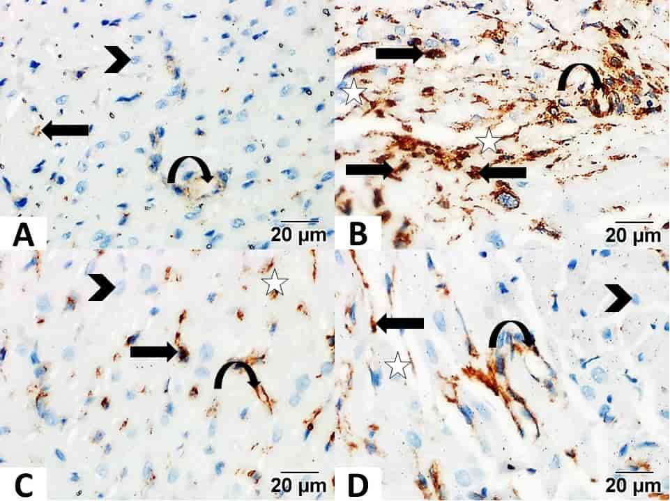
Fig.1: Photomicrograph of control showed minimal immune reaction in the blood capillaries wall (curved arrows) and interstitial cells (arrow). With an immune negative cardiac muscle fibers (arrowhead). (B). DOX group showed strong immune reaction in endomysium and perimysium connective tissues (star), in the blood capillaries wall (curved arrows), and interstitial cells (arrow). (C). DOX + CoQ10 showed mild immune reaction in the endomysium and perimysium (star), in the blood capillaries wall (curved arrows) and interstitial cells (arrow). With an immune negative reaction in cardiac muscle fibers (arrowhead). (D): DOX + L-carnitine showed moderate immune reaction (Vimentin. Scale bar 20µm).
Mahtab Rahbar
Iran University School of Medicine, Iran
Title: Histopathology review of idiopathic steroid resistant nephrotic syndrome and outcome in children in north-west of Iran

Biography:
Mahtab Rahbar is pathologist from Tehran medical university .she got dermatopathology fellowship from Tehran medical university also. Currently, she is associated professor and faculty member for 25 years. She is the director of clinic pathology department of Ali Asghar hospital. She has published more than 35 papers in reputed journals and has been serving as an editorial board member of repute.
Abstract:
There is currently little information in the literature on the spectrum of histopathologic patterns in children presenting with idiopathic steroid-resistant nephrotic syndrome (iSRNS) in Iran. We conducted to compare the histopathologic distribution of different subtypes’ glomerular morphologic patterns in iSRNS and the clinical and biochemical parameters at the time of diagnosis and outcome of patients after immunosuppressive therapy. Material and Methods: This cross sectional study was done in two hundred children, aged 1 - 15 years, who were diagnosed for iSRNS and no response to 4 weeks of standard prednisone therapy (60 mg/m2/day) referred to nephropathology Department of Emam Reza hospital between 2005 and 2013. Demographic, clinical, laboratory, and histopathological data were retrieved from files and original renal biopsy reports. We discussed histopathologic diagnosis and outcome of iSRNS after initial therapy in patients separately. This study investigated prognostic effects of histopathologic pattern on outcome of iSRNS. Results: The study included 200 children with iSRNS: 141 (70.5%) were males and 59 (29.5%) females, with male-to-female ratio of 2.4:1. The mean age was 7.23 ± 4.37 years (range: 1 - 15 years). Upon pathologic investigation of iSRNS cases, focal segmental glomerulosclerosis (NOS subtype) was the first, with a highest prevalence at a rate of 102/200 (51%) and MGN was the last, at a rate of 7/200 (3.5%). Children with iSRNS secondary to MCD are more likely to achieve remission and have better long term prognostic value (P < 0.00). Focal segmental glomerulosclerosis (FSGS) (Tip and Collapse subtypes) is more likely to have worse outcome in response to immunosuppressive therapy (P < 0.04). Conclusions: This study shows that the response to cyclosporine can be correlated with the underlying histopathology patterns which have been earned by adequate renal biopsy.
Biography:
John Lucci has completed his MS at the age of 25 years from University of Rhode Island. He has rotated through eight medical centers in New England. He has written almost 100 papers, available online through open access and has posted more than 80 clinical cases on social media.
Abstract:
A review of 22 cases of metastases to Cerebrospinal Fluid (CSF) in a non-pediatric population from 20 different primary sites as catalogued at multiple medical centers in Boston. Clinical correlation with outcome is detailed, along with morphological charicteristics specific to metastases in CNS fluids.
Qamar-un-Nisa
University of Veterinary and Animal Sciences, Lahore, Pakistan
Title: Pathological alterations during co-infection of newcastle disease virus with Escherichia coli in broiler chicken

Biography:
Dr.Qamar-un-Nisa, currently serving as Faculty member in Department of Pathology, Faculty of Veterinary Science, University of Veterinary & Animal Sciences, (UVAS) Lahore.. For Higher studies, Dr.Qamar un Nisa has been awarded indigenous Ph.D scholarship funded by Higher Education Commission(HEC)Government of Pakistan. She has over 10 years teaching and research experience. She is involved in teaching, research, laboratory Work and Postmortem examination of large and small animals.She also deals with veterolegal cases. She is the member of board of studies of Pathology Department. She has attended many conferences and seminars nationally and internationally. She has also attended 5th Turkish Vet. Pathology Congress, Bursa-Turkey and orally presented her research paper on ‘’Histopathological and hematological analysis of broiler chicken experimentally infected with pathogenic E.coli.’’ Dr.Qamar-un-Nisa has over 15 research publications in well reputed national and international journals.Her areas of interest are Histopathology , Cytopathology and Diagnostic Pathology
Abstract:
Respiratory diseases are responsible for major economic losses at poultry farms especially during co-infections of respiratory pathogens. However, impact of co-infections is not well known, especially in broilers. The current study was aimed to assess the probable synergism of E. coli (O78) and velogenic Newcastle disease virus (vNDV-CK-Pakistan-NARC-13N39-2013), in the broiler model. Three-week-old commercial broilers were inoculated with either vNDV, E. coli serotype O78 or both agents simultaneously or 3 days apart. The birds were clinically observed and swabbed daily. They were killed at 4 and 14 days after single or dual inoculations and were inspected for gross lesions. Samples of the respiratory organs (trachea, lungs, and air sacs) were taken for histological analyses. All the infected subjects showed clinical signs of varying severity. Co-infected groups showed the most obvious clinical signs, associated with significant higher mortality and respiratory organ abnormalities, in comparison with the monoinfected groups (P<0.05). There was a non-significant (P>0.05) effect of the inoculation time intervals between vNDV and E. coli inoculation (none or 3 days). Microscopic lesions staining supported clinical and macroscopic findings. Higher virus shedding (P<0.05) in oropharyngeal swabs was observed in coinfected groups than single infected groups. The results revealed that experimental co-infection of E. coli and NDV enhances the degree of severity of clinical signs, gross lesions and death rate and warns that E. coli and NDV can cause substantial economic losses by exercising additive or synergistic pathogenic effect in the reproduction of respiratory disease if given simultaneously or three days apart.
Zhang Jinxia
Shougang Hospital of Beijing University, China
Title: Potential antigen targets profiling for colorectal cancer immunotherapy

Biography:
Abstract:
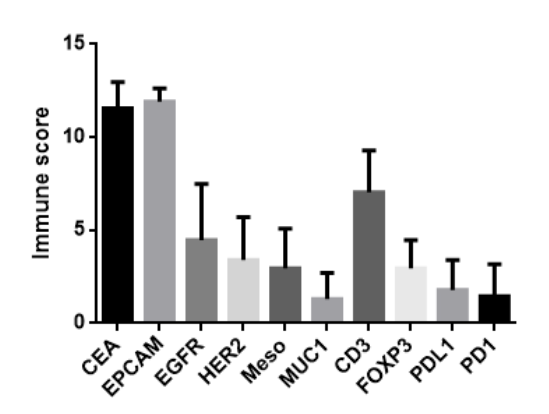
Background: Adoptive chimeric antigen receptor T (CAR-T) cells and immune checkpoint inhibitors have been proven to be the promising therapies for the treatment of solid tumors in recent years. However, the cell surface antigen expression plays a vital role in both of these immunotherapies. In this study, we investigated the expression of 6 cancer-associated antigens( epithelial cell adhesion molecule (EpCAM); carcinoembryonic antigen(CEA); epidermal growth factor receptor(EGFR); mesothelin;mucin 1(MUC1); epidermal growth factor receptor 2(HER2)) and 4 immune microenvironments associated markers(CD3 ; programmed death 1(PD1); programmed death-ligand 1 ( PDL1) ; forkhead box P3(FOXP3))in colorectal cancer.
Methods: All available formalin-fixed, paraffin-embedded tumor slides from 113 colorectal patients were reviewed. Intensity and distribution for each antigen were assessed by immunohistochemistry.
Results: Positive expression of EpCAM, CEA, EGFR, Mesothelin, MUC1, and HER2 were demonstrated in100%, 99%, 96%, 68%, 67% and 37% of colorectal cancer respectively. As for immune microenvironment, CD3, PD1, PDL1 and FOXP3 were positive in 100%, 73%, 71%,97% colorectal cancer cases. More than 90% cases had≧75% distribution of EpCAM-positive cells and CEA-positive cells. More than 50% cases had≧50% distribution of CD3-positive cells, in which almost 40% CD3 positive cells were also FOXP3 positive. However, the PD1 and PDL1 expression were very low in colorectal cancer.
Conclusion: EpCAM and CEA expression were very high in colorectal cancer, which could be the potentially promising targets for colorectal cancer CAR T cell therapy. Although PD1 and PDL1 expression were low in colorectal cancer microenvironment, it could be another strategy by targeting regulatory T cells (FOXP3 positive) to relieve the immunosuppression and enhance the antitumor function of the immune system for colorectal cancer patients.
Figure1. The expression of 6 cancer-associated antigens and 4 immune microenvironment associated markers in colorectal cancer.
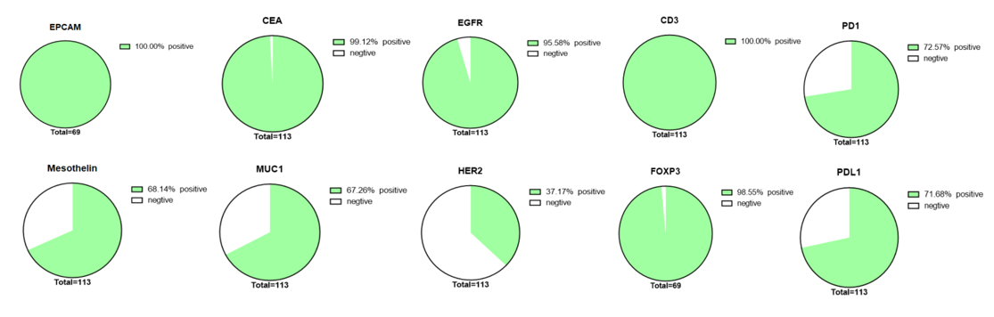
Figure2. The immune scores of 6 cancer-associated antigens and 4 immune microenvironment associated markers in colorectal cancer.
Asmaa Elsayed Bedeer Mohammed
Tanta University, Egypt
Title: Significance of tissue expression and serum levels of angiopoietin-like protein 4 in breast cancer progression: Link to NF-κb /P65 activity and pro-inflammatory cytokines

Biography:
Asmaa Elsayed Bedeer Mohammed, lecturer of histopathology, faculty of medicine, Tanta University.
Abstract:
Background: The molecular mechanisms linking breast cancer progression and inflammation still remain obscure. The aim of the present study was to investigate the possible association of angiopoeitin like protein 4 (ANGPTL4) and its regulatory factor, hypoxia inducible factor-1 α (HIF-1α), with the inflammatory markers nuclear factor kappa B/p65 (NF-κB /P65) and interleukin-1 beta (IL-1β) in order to evaluate their role in inflammation associated breast cancer progression.
Materials and Methods: Angiopoietin-like protein 4 (ANGPTL4) mRNA expressions were evaluated using quantitative real time PCR and its protein expression by immunohistochemistry. DNA binding activity of NF-κB /P65 was evaluated by transcription factor binding immunoassay. Serum levels of ANGPTL4, HIF-1α and IL-1β were immunoassayed. Tumor clinico-pathological features were investigated.
Results: ANGPTL4 mRNA expressions and serum levels were significantly higher in high grade breast carcinoma (1.47±0.31 and 184.98±18.18, respectively) compared to low grade carcinoma (1.21±0.32 and 171.76±7.58, respectively) and controls (0.70±0.02 and 65.34±6.41, respectively), (p<0.05). Also, ANGPTL4 high/moderate protein expression was positively correlated with tumor clinico-pathological features. In addition, serum levels of HIF-1α and IL-1β as well as NF-κB /P65 DNA binding activity were significantly higher in high grade breast carcinoma (148.54±14.20, 0.79±0.03 and 247.13±44.35 respectively) than their values in low grade carcinoma ( 139.14±5.83, 0.34±0.02 and 184.23±37.75, respectively) and controls (33.95±3.11, 0.11±0.02 and 7.83±0.92, respectively), (p<0.001).
Conclusion: ANGPTL4 high serum levels and tissue expressions in advanced grade breast cancer, in addition to its positive correlation with tumor clinico-pathological features and HIF-1α could highlight its role as one of the signaling factors involved in breast cancer progression. Moreover, novel correlations were found between ANGPTL4 and the inflammatory markers, IL-1β and NF-κB/p65, in breast cancer, which may emphasize the utility of these markers as potential tools for understanding interactions for axes of carcinogenesis and inflammation contributed for cancer progression. It is thus hoped that the findings reported here would assist in the development of new breast cancer management strategies that would promote patients’ quality of life and ultimately improve clinical outcomes. However, large-scale studies are needed to verify these results.
Shankar Bastakoti
BP Koirala Memorial Cancer Hospital, Nepal
Title: Fine needle aspiration cytology in head and neck lesions: A tertiary hospital based experience in eastern Nepal

Biography:
Registrar Pathologist BP Koirala Memorial Cancer Hospital, Bharatpur, Nepal
Abstract:
Fine Needle Aspiration Cytology (FNAC), a simple and rapid diagnostic technique being considered as a valuable diagnostic aid because of the early availability of results, simplicity, easy accessibility, minimal trauma and absence of complications.
AIMS AND OBJECTIVES:
Study the cytological spectrum and systemic analysis of the head and neck lesions and correlate with histopathologic diagnosis. And evaluate the sensitivity, specificity & diagnostic accuracy of the fnac.
Hospital based Cross-Sectional study, was conducted in the Department of Pathology at BPKIHS, Dharan.
Our study included 404 cases with the female: male ratio 1.6:1. Overall benign lesion (38.6%) outnumbered the malignant lesion (21.3%). Female presented with thyroid lesion (43.1%) commonest likewise lymphnode (52.5%) in a male. Altogether 69 cases were diagnosed malignant and 17 were suspicious for malignancy however only 29 could be correlated histopathologically. EIC as benign and Squamous Cell Carcinoma as malignant lesion are the most frequent in Cutaneous and soft tissue. Likewise, Colloid Nodule, Papillary carcinoma and lymphocytic thyroiditis in inflammatory lesion of thyroid. Pleomorphic adenoma, Mucoepidermoid carcinoma, and chronic sialadenitis observed most in salivary gland. Reactive and granulomatous lymphadenitis were the commonest in lymphnode and Malignant lesion were observed comprising of Metastatic Carcinoma and hematolymphoid which was the commonest site for malignancy. The overall sensitivity, specificity and diagnostic accuracy was 96.5%, 98.4% and 97.8%.
FNAC in our experience proved effective, efficient, prompt diagnosis and minimal invasive procedure hence recommended as the first line investigation in case of any head and neck lump/mass/lesion.
Preksha Sharma
S.M.S Medical College, Jaipur, India.
Title: Umbilical cord- a key factor in still births and full term normal deliveries

Biography:
M.B.B.S, M.S. (Anatomy) S.M.S Medical College, Jaipur, Rajasthan, India.
Abstract:
INTRODUCTION:
Stillbirth is defined as “when the infant is delivered with no signs of life and the gestational age being between 20 weeks and full term”. It is one of the obstetric complications which have not been studied widely. Still birth presents a devastating pregnancy outcome and the need for increased efforts in prevention has been highlighted. The umbilical cord, as documented in the literature is a cord like structure covered by the amniotic membrane. The fetal well-being is adversely affected by the pathologic lesions of the umbilical cord. There are numerous umbilical cord abnormalities, both gross and histological which have been associated with still birth. Compromise of foetal umbilical circulation is seen in 20% of stillbirths at the time of autopsy.
MATERIAL AND METHOD:
In the present study the cases were divided into two groups i.e. group A (control), group B (cases of still births) and parameters were record
RESULT:
GROSS EXAMINATION OF UMBILICAL CORD
- Length of the umbilical cord
Length of umbilical cord in normal full term deliveries is significantly more (p<0.05) as compared to that of the still births
- Knots in the umbilical cord
The difference in proportion of false knot was not with significant (p>0.05) variation. True knots were not found.
- Insertion of the umbilical cord
Insertion was found to be central in both the groups
HISTOLOGY OF UMBILICAL CORD
- UMBILICAL CORD VASCULITIS
Vasculitis was seen only in the cases of still birth.
- INFLAMMATORY CELLS
None in Group A whereas in group B there were inflammatory cells in umbilical cord in 16%.
CONCLUSION:
There were increased inflammatory changes such as vasculitis and inflammatory cell infiltrate in the case of still birth. Therefore appropriate and timely measures should be taken in an order to improve the outcome of pregnancy.
Odukoya Lateef A
Lagos University Teaching Hospital, Lagos Nigeria
Title: Histopathologic patterns of salivary gland tumors (sgts): A 10-year retrospective study

Biography:
Odukoya Lateef A is a senior registrar at the department of anatomic and molecular pathology of the Lagos University Teaching Hospital. He is a young pathology trainee with an interest in cancer research and health promotion. He looks forward to further trainings/exposures and collaborations in the areas of cancer research.
Abstract:
The incidence of salivary gland tumors is influenced by geographical and racial factors. The histopathologic classification and nomenclature of salivary gland neoplasms as defined by WHO classification (2005), is accepted globally but little is available in the literature regarding the trend of these tumors in Africa based on this classification. Our study was to outline the histopathologic patterns of SGTs in a Tertiary center in Lagos Nigeria.
This was a retrospective study; data was retrieved from the records at the department of Anatomic and Molecular Pathology and Oral pathology at the Lagos University Teaching Hospital from 2006 to 2015. All SGTs were grouped using the WHO 2005 Classification of SGTs.
172 cases of salivary gland neoplasms were diagnosed. There were 88(51.1%) males and 84(48.6%) females. The age range of the 170 patients with recorded ages was from 4 to 85 years. The mean age at diagnosis was 43.5 years (SD =59.2 years) and median was 40.8 years.
44.2% of neoplasms affected the parotid, 19.8% involved the submandibular and 34.9% affected the MiSGs. Malignant tumors occurred more in the MiSGs (47.9%). Malignant benign tumors accounted for 54.7% and 45.3% respectively. Pleomorphic adenoma was the most common benign tumor (94.8%), followed by basal cell adenoma (3.9%). No Whartin’s tumor was found. The malignant tumors was dominated by adenoid cystic carcinoma (40.4%) followed by mucoepidermoid carcinoma (29.8%).
Overall the patterns seen in our study corresponds with many the previous researchers. In that we a dominance of adenoid cystic carcinoma as well as predilection of malignant tumors for the minor glands.
Deva Japa
Gujarat Methodist Church Cardiac Care Society, India
Title: Giant left atrial myxoma induces mitral valve obstruction and pulmonary hypertension

Biography:
Dr. Deva Japa Ajith, MD –Pathology, now is a Senior Research Scientist , Gujarat Methodist Church Cardiothoracic and Vascular Research Society, Consultant Pathologist, Department of Pathology , Dharamsinh Desai Methodist Memorial Cardiac Institute , Transfusion Officer- Department of Blood Banking , Gujarat Methodist Church Cardiac Care Society,India and Visiting Faculty in the Charotar University. Done M.B.B.S from Kilpauk Medical College, Chennai- India, Internship -Internal Medicine, Kilpauk Medical College, Chennai, India and MD from Christian Medical College, Ludhiana, India. Positioned as supervisor for many Postgraduate Biomedical and Biotechnology graduates in their dissertation. Member,Institutional Review Board,Research Society, DharamSinh Desai Methodist Mission,Nadiad,Gujarat,India , Board of Committee member in the state Blood Transfusion council and member of Indian Society of Pathology and Microbiology
Abstract:
Primary intracardiac tumour in the site of left ventricle was described in 1559 and myxoma seems to constitute 40-50% of the primary intracardiac tumours. Atrial myxomas are sporadic in origin with unknown aetiology and nearly 90% appear to be solitary and pedunculated;75-85% seen in left atrial cavity. It is often attached to the atrial septum. It occurs in middle age with mean age of 56 years and more common in women. It could be familial in up to 10% of cases and when this occur, they are more likely to be multiple and located in the ventricle also.
The symptoms of atrial myxoma are related to location, embolization and propensity to obstruct blood flow through the heart and are clinically difficult to diagnose. Diagnosis was possible only at post-mortem till 1951,after which diagnosis of left atrial tumour has been confirmed by angiocardiography. Till today echocardiography is a tool to diagnose intracardiac masses and it poses challenges in discrimination between primary cardiac tumours, such as myxoma, and other cardiac masses; which can be specified by histopathological examination. As literature reveals there is low surgical mortality and long term good results in patients with cardiac myxoma.
Histologically presence of myxoma cells in the background of myxoid stroma is the diagnostic point. But the most challenging differential diagnosis is with mural thrombi showing myxoid changes. Calretinin is a novel marker which is positive in the myxoma cells and hence helps in differentiation from mural myxoid thrombi. Positive expression of Calretinin marker indicate that the myxoma cells may originate from endocardial sensory nerve tissue.
In conclusion, Giant Left Atrial Myxoma is a rare benign lesion of the heart which can be treated by surgical removal. In general, it appears small and asymptomatic. However, it may present as large mass rarely because of the asymptomatic course despite the large size of the tumour and clinically mimic malignancy. Embolization of its fragments may cause pulmonary embolism when located in the right and systemic organ infarction when located on the left. Interference with mitral flow mimic mitral stenosis with massively dilated left atrium, pulmonary hypertension and chronic heart failure which progress over an appreciable duration of time.
DEMANOU KENGNI Georges Gonstran
University Evangelical Institute of Cameroon, BP
Title: Evaluation of the quality of cervico-vaginal smears at gyneco-obstetric and pediatric hospital of douala

Biography:
Department of Biomedical Sciences, Faculty of Science and Technology, University Evangelical Institute of Cameroon
Abstract:
Introduction: Cervical cancer is likely to be cancer whose Cervico-Vaginal Smear (CVF) screening would be the most contributive if performed under favorable conditions. In Cameroon, the laboratories of Anatomy Cytology Pathology (ACP) are very few, there is no quality control in these laboratories and even less, there is no study on the quality of samples of CVS since its implementation in our country. However, in the laboratory, the quality of the sample determines the quality of the analysis and consequently the quality of the result and therapeutic orientations. Thus, the quality and safety of CVS results could be challenged by poor sample quality. The objective of our study was to evaluate the quality approach in Cervico-Vaginal smears, recognized as the examination of choice in the early detection of precancerous lesions of the cervix.
Methods: We conducted over a period of 16 months (September 2015 to December 2016), a retrospective study of descriptive type over a period of 08 months (December 2016 to July 2017) at the Gyneco-Obstetrics and Pediatric Hospital of Douala where we have performed a quality control of Cervico-Vaginal smears quality criteria only at the sampling level using the Bethesda System as reference quality manual.
Results: A total of 508 CVS in clinical practice were reviewed and at the end of the analyzes, we obtained 28.94% CVS satisfactory for evaluation, 43.31% CVS satisfactory but limited by ... and the CVS rate was inadequate 27.76%. The analysis of the reports revealed that 65.31% of the CVS had no information regarding the quality of the sample.
Conclusion: Given these results, the quality of samples taken at HGOPED is partially in line with Bethesda's recommendations, which implies that ACP professionals must really engage in a process of quality improvement in the practice of CVS.
Thanaa El Sayed Ahmed Helal
Ain Shams University, Egypt
Title: Immunohistochemical and molecular profile of epithelial mesenchymal transition markers in hepatocellular carcinoma patients in Egypt

Biography:
Thanaa El Sayed Ahmed Helal is a professor emeritus in pathology, faculty of medicine, Ain Shams University. Her expertise in evaluation and interpretation pathology repors, immunohistochemistry techniques and frozen section diagnosis is high reputated. She participated in establishing the Egyptian committee for training pathologists with conjunction with the Royal Collage of Pathologists, UK. Her expertise expands in teaching, diagnosis and research. Her research implies a variety of techniques and multidiscipline. In the last decade, she focused on liver pathology research. She has been granted several national awards and national grants for funding research projects. She is a member of many national and international pathology societies, member of the Supreme Council of universities for promoting pathology professors.
Abstract:
Statement of the Problem: The currently used clinical and pathologic prognostic parameters are not fully valid in all hepatocellular carcinoma (HCC) cases, since HCC is highly heterogeneous and tumors with similar clinical and pathological features may behave differently. The achievement of comprehensive molecular classification of HCC may hopefully help in identifying valuable therapeutic targets. The role of epithelial mesenchymal transition (EMT) markers has been investigated in various cancers with few studies on HCC and no reports are available from Egyptian HCC patients. The purpose of this study is to identify novel tissue biomarkers that can be used as reliable prognostic factors to predict recurrence and metastasis in HCC patients. Methodology & Theoretical Orientation: HCC liver tissue specimens from 100 patients, who underwent hepatectomy with available follow-up data, were immunostained with E-Cadherin, Keratin, Vimentin, N-Cadherin, Integrins, CDX2, Stat-3, SNAIL, Slug, Zeb1and Twist. Real-time PCR was performed to analyze the molecular profile of EMT. Results of immunostaining were correlated with the clinicopathological features, molecular profile as well as the follow-up data. Findings: Upregulation of Keratin, Vimentin and E-Cadherin expression profile were significantly correlated with extrahepatic recurrence after 12 months following curative surgery (p<0.001, p=0.001, p<0.001) and after 24 months (p<0.001, <0.001, <0.001) respectively. Moreover, Keratin, Vimentin and E-Cadherin expression profile were significantly correlated with microvascular invasion (p=0.015, p=0.015, p=0.11) respectively. None of N-Cadherin, Integrin, CDX2, STAT3, Zeb1, Twist, SNAIL and Slug were correlated with extrahepatic recurrence following curative surgery or microvascular invasion. No correlation was detected between any of the 11 EMT markers and AFP level, advanced AJCC tumor stage or tumor histological grade. Conclusion & Significance: EMT expression profiles are useful prognostic markers for recurrence in HCC patients. High Keratin, Vimentin and E-Cadherin expression profile is closely associated with advanced tumor stage, microvascular invasion and metastasis indicating poor tumor behavior.

Biography:
Abstract:
INTRODUCTION:Â Â Â
Liquid bases cytology (LBC) has become increasingly popular in gynaecological pathology, however, it is gaining importance in the evaluation of non-gynaecologic cytology specimens including fine needle aspiration (FNA). The more widely used technologies for liquid-based cytology require expensive equipment, hence the need to evaluate and validate a cheaper & inexpensive manual method for processing LBC specimens.
AIMS AND OBJECTIVES:
The aims and objectives of the study was to evaluate the efficacy of manual LBC in FNA cytology versus conventional slide preparation method, study cytomorphological features of various lesions on liquid-based preparation and assess the sensitivity and specificity of the diagnosis rendered on liquid based preparation wherever possible.
MATERIALS AND METHODS:
FNA was performed on 50 cases with superficial palpable swellings. Material was obtained by minimum of 3 passes in each case for conventional and manual liquid-based preparation of aspirates using SurePathTM cytokit.
RESULTS:
Out of 50 total cases, 20 were performed on thyroid swelling, 14 on lymphadenopathies and 16 on palpable breast lumps. Manual LBC had sufficient cellular yield with 98% cases depicting adequate material for diagnosis. Histopathological confirmation was available in 29 of 49 reported cases with a 100% sensitivity and specificity. LBC has shown superiority in terms of cellularity, cell arrangement, cellular details, background and reduced screening time and storage.
CONCLUSION:
Manual LBC gives superior results when compared with that of the conventional method with better morphology. Various artifacts inherent to liquid-based cytology should be known and kept in mind during reporting.Â